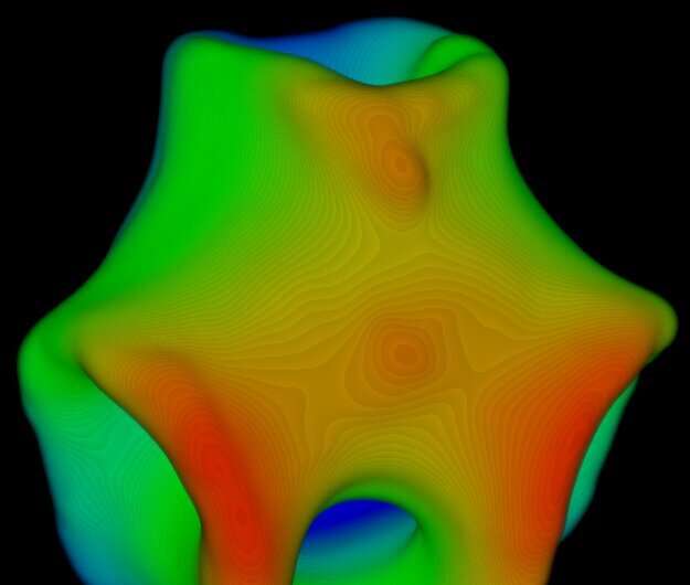
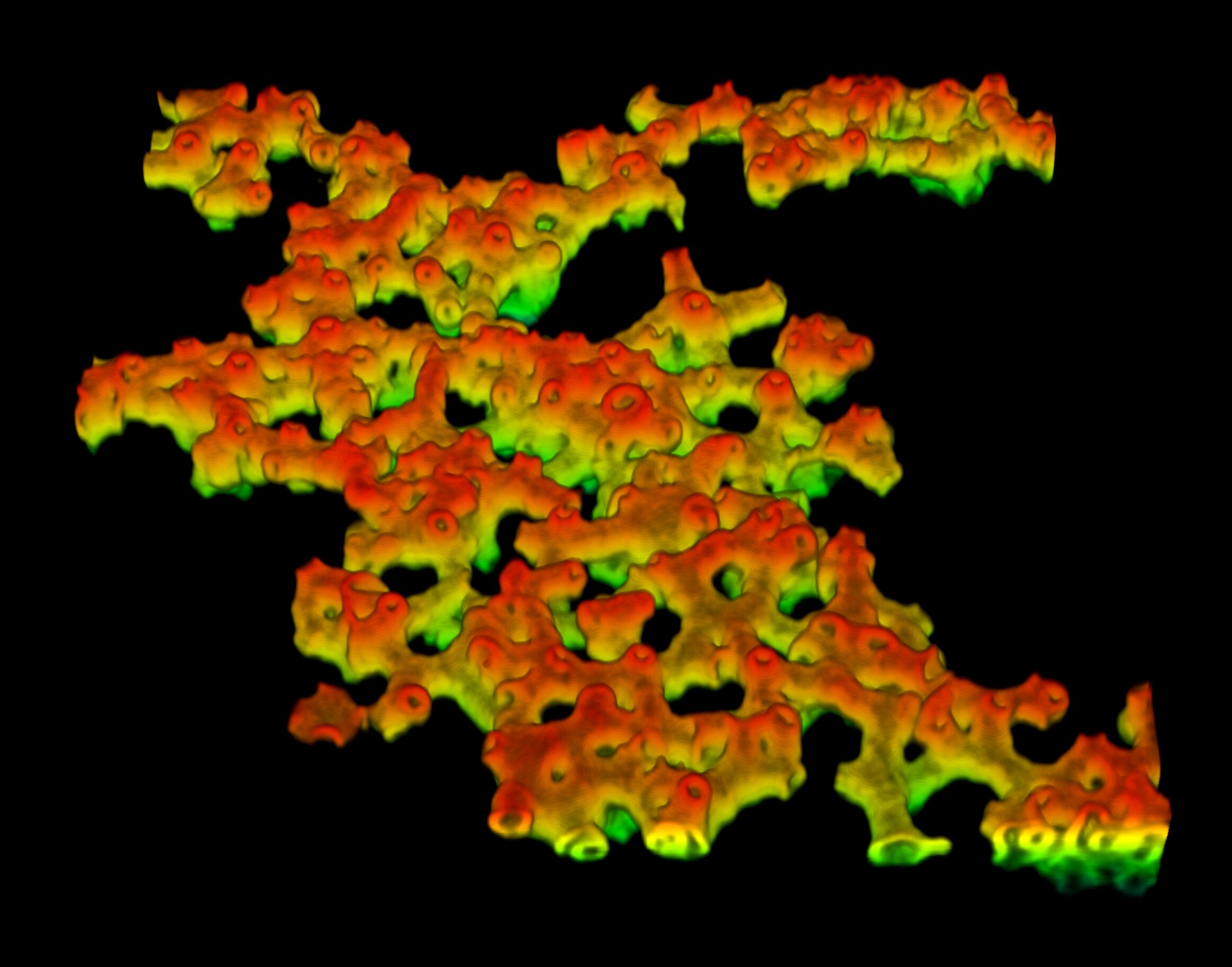
Image (false colors) of a spongy phase of fluid colloidal membranes, which are themselves composed of a binary mixture of short and long rods. Photo credit: Ayantika Khanra
Cell membranes transition seamlessly between different 3D configurations. It is a remarkable feature that is essential for several biological phenomena such as cell division, cell mobility, transport of nutrients in cells, and viral infections. Researchers at the Indian Institute of Science (IISc) and their collaborators recently devised an experiment that sheds light on the mechanism by which such processes might occur in real time.
Researchers studied colloidal membranes, which are micron-thick layers of aligned, rod-shaped particles. Colloidal membranes offer an easier system to study because they share many of the same properties as cell membranes. Unlike a sheet of plastic, where all molecules are immobile, cell membranes are liquid sheets in which each component is free to diffuse. “This is a key property of cell membranes that is available in ours [colloidal membrane] system,” explains Prerna Sharma, associate professor in the Institute of Physics at IISc and corresponding author of the study published in the journal Proceedings of the National Academy of Sciences.
The colloidal membranes were assembled by preparing a solution of rod-shaped viruses of two different lengths: 1.2 microns and 0.88 microns. The researchers studied how the shape of the colloidal membranes changes as you increase the proportion of short rods in the solution. “I made multiple samples by mixing different volumes of the two viruses and then observing them under a microscope,” explains Ayantika Khanra, Ph.D. Student at the Institute of Physics and first author of the work.

Image (false colors) of a fluidic colloidal membrane that is itself composed of a binary mixture of short and long rods. Photo credit: Ayantika Khanra
When the proportion of short rods was increased from 15% to 20-35%, the membranes transitioned from a flat disk-like shape to a saddle-like shape. Over time, the membranes began to fuse together and increase in size. Saddles have been classified by their order, which is the number of ups and downs encountered as you move along the edge of the saddle. The researchers observed that the laterally converging saddles formed a larger saddle of the same or higher order. However, when they merged at an almost right angle away from their edges, the final configuration was a catenoid-like shape. The catenoids then merged with other saddles, creating increasingly complex structures such as trinoids and quad-noids.
To explain the observed behavior of the membranes, the researchers also proposed a theoretical model. According to the laws of thermodynamics, all physical systems tend to move toward low-energy configurations. For example, a drop of water takes on a spherical shape because it has less energy. For membranes, this means that shapes with shorter edges, such as a flat disk, are preferred. Another property that plays a role in defining the membrane configuration is the Gaussian curvature modulus. A key finding of the study was to show that the Gaussian curvature modulus of the membranes increases as the fraction of short rods is increased. This explains why adding more short rods drove the membranes to saddle-like shapes that have less energy. It also explains another observation from their experiment where lower-order membranes were small while higher-order membranes were large.
“We have proposed a novel mechanism for curvature generation of fluid membranes. This curvature adjustment mechanism by changing the Gaussian modulus could also play a role in biological membranes,” says Sharma. She adds that they plan to further investigate how other microscopic changes in membrane components affect the large-scale properties of membranes.
Burning membranes for molecular sieving
Ayantika Khanra et al, Controlling the shape and topology of two-component colloidal membranes, Proceedings of the National Academy of Sciences (2022). DOI: 10.1073/pnas.2204453119
Provided by the Indian Institute of Science
Citation: 3D shaping of microscopic membranes underlying cellular processes (2022 September 12) retrieved September 12, 2022 from https://phys.org/news/2022-09-3d-microscopic-membranes-underlie- cellular.html
This document is protected by copyright. Except for fair trade for the purpose of private study or research, no part may be reproduced without written permission. The content is for informational purposes only.
#shaping #microscopic #membranes #underlying #cellular #processes


Leave a Comment