Summary: CSF drainage pathways are similar in mice and humans, researchers discovered.
Source: jale
Meningeal lymphatics are potential targets for treating brain diseases. Laboratories at Yale and the Paris Brain Institute (Pitié-Salpêtrière Hospital, Paris) have imaged brain drainage through meningeal lymphatics in mice and humans.
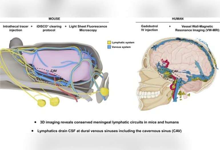
The recent Journal of Experimental Medicine Paper led by Jean-Leon Thomas, Ph.D., professor of neurology, and Anne Eichmann, Ph.D., ensign professor of medicine and professor of cellular and molecular physiology and co-director of the Cardiovascular Research Center (YCVRC ) at Yale, shows that CSF drainage pathways are similar between mice and humans, and reports a novel MRI-based imaging technique for patients with neurological disorders.
The lymphatic vasculature controls immune surveillance and waste clearance in tissues and organs. Lymphatic vessels are absent from the central nervous system (CNS) but are present at the CNS borders, in the meninges that protect the brain and spinal cord. The meningeal lymphatics drain into the cervical and peripheral immune system lymph nodes, making them key players in controlling brain immunity.
The meningeal lymphatics are also important in the removal of waste from the brain, by being involved in the removal of interstitial fluid and soluble proteins, and in the drainage of cerebrospinal fluid, which provides the brain with a protective fluid buffer against injury, a pathway for essential nutrients and Disposal system for cell waste.
The meningeal lymphatic system affects neurological diseases in many mouse models, including Alzheimer’s disease, multiple sclerosis, brain tumors, and other diseases. “Due to its involvement in many diseases, the meningeal lymphatic system has attracted a great deal of therapeutic interest,” explains Laurent Jacob, Ph.D., first author of the study and member of the Paris research team.
“However, it remained unclear where the lymphatic reuptake of CSF molecules occurs in the context of the whole head, in mice or in humans.”
To learn more about the architecture and function of the meningeal lymphatic network, the team studied CSF lymphatic drainage using postmortem light sheet imaging in mice and real-time magnetic resonance imaging in humans. By combining these approaches, the authors rebuilt the entire CSF lymphatic drainage network.
3D imaging showed that the meningeal lymphatics contact the venous sinuses of the dura mater and revealed an extensive meningeal lymphatic network around the cavernous sinus in the anterior aspect of the skull. From there, meningeal lymphatics leave the skull through cranial foramina and drain into cervical lymph nodes.
Stéphanie Lenck, MD, also at Pitié-Salpêtrière Hospital, performed quantitative lymphatic MRI on 11 patients with various neurological disorders. She established a method for the 3D visualization of all blood and lymphatic vessels in the meninges and neck, which showed a significantly larger meningeal lymph volume in men than in women.
Whether these anatomical data are causally related to the higher predisposition of women to develop neurological diseases such as multiple sclerosis, meningiomas or intracranial hypertension needs to be researched in the future.
“Meningeal lymph vessels are potential targets for the treatment of brain diseases,” says Eichmann. “Labs at Yale are making strides in elucidating their function by imaging brain drainage through meningeal lymphatics in mice and humans.”
About this news from neuroscientific research
Author: Elizabeth Reitman
Source: jale
Contact: Elizabeth Reitman-Yale
Picture: The image is credited to the researchers
See also

Original research: Open access.
“Preserved meningeal lymphatic drainage circuits in mice and humans” by Laurent Jacob et al. Journal of Experimental Medicine
abstract
Preserved meningeal lymphatic drainage circuits in mice and humans
Meningeal lymphatic vessels (MLVs) have been identified in the dorsal and caudobasal regions of the dura mater, where they provide waste clearance and immune surveillance of brain tissues. Whether MLVs exist in the anterior part of the murine and human skull and how they are associated with the glymphatic system and extracranial lymphatics remained unclear.
Here we used light sheet fluorescence microscopy (LSFM) imaging of mouse whole head preparations after OVA-A555 Cerebrospinal fluid (CSF) tracer injection and real-time magnetic resonance imaging (VW-MRI) of the vascular wall (VW) following systemic injection of gadobutrol in patients with neurological pathologies.
We observed a conserved three-dimensional anatomy of MLVs in mice and humans, consistent with dural venous sinuses but not with nasal CSF outflow, and we discovered an enlarged anterior MLV network around the cavernous sinus with exit pathways through the foramina of the emissary veins. VW MRI can be a diagnostic tool for patients with cerebrospinal fluid drainage defects and neurological disorders.
#brains #drainage #system #Neuroscience #News


Leave a Comment