Steatocystoma multiplex is a rare condition consisting of multiple cysts erupting over the chest, arms, armpits and neck and is usually inherited in an autosomal dominant manner [1]. These lesions are largely asymptomatic but may erupt and secrete an oily substance if the cyst is near the skin’s surface [2]. Many of the features of this condition overlap with a variety of diseases such as eruptive vellus hair cyst (EVHC) and acne vulgaris. A differentiation is therefore important, since the clarification and treatment is very different [2,3]. This case report outlines a presentation of steatocystoma multiplex to draw attention to the unique clinical features of this rare disease.
A 47-year-old woman with a history significant only for recurrent skin infections presented for a routine mammography without symptoms or concerns.
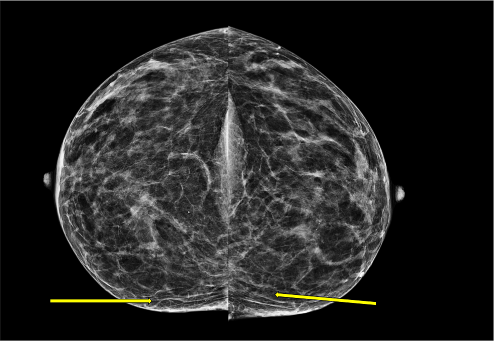
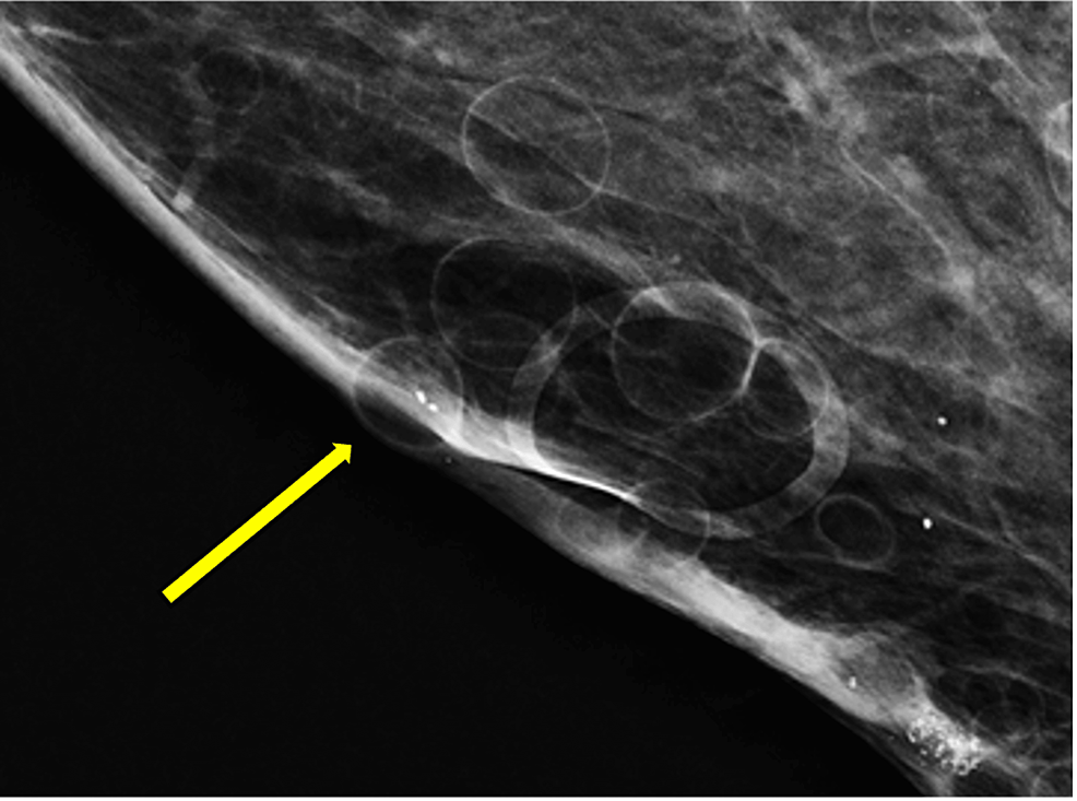
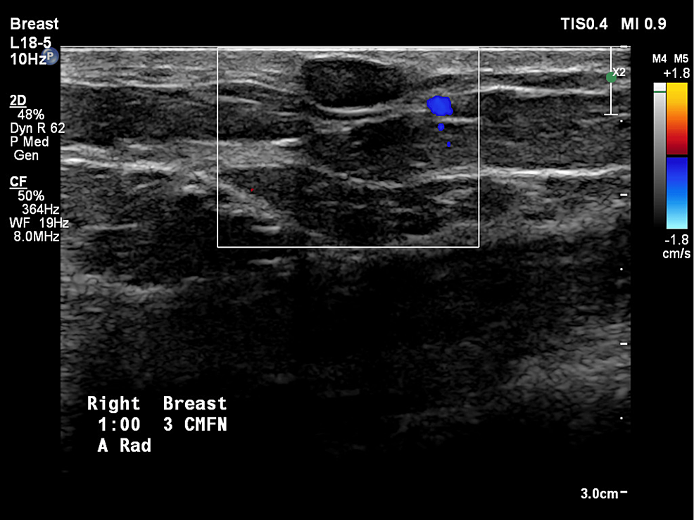
The patient’s screening mammography was performed (Figures 1, 2) and innumerable oil cysts in both breasts, predominantly medial. The patient had a diagnostic mammogram with targeted bilateral breast ultrasound that revealed multiple oval anechoic cystic masses consistent with the mammographic findings of multiple oil cysts. A follow-up ultrasound was performed (Fig 3) showing a circumscribed, avascular oil cyst.
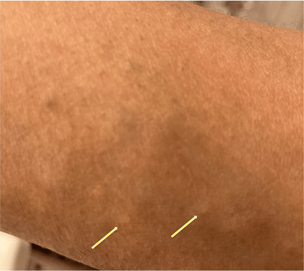
Upon examination of the patient’s medical record, she had no history of breast injury, previous breast surgery, or breast discomfort. The patient was contacted following her imaging results. She denied any significant medical history or family history of breast or skin pathologies at the time. She noted that since early adulthood she had consistently had multiple “bumps” on her torso, chest, and inner arms, which represented subcutaneous cysts, the number of which slowly increased over time (Fig 4). She also notes a subcutaneous cyst on her chest that occasionally ruptures and bleeds oily fluid. She denied any other associated symptoms or concerns about her cysts, saying she was the only one in her family with these symptoms.
The patient’s GP has been contacted regarding the findings and will continue to monitor her symptoms on an outpatient basis.
Steatocystoma multiplex is a rare benign condition that overlaps clinical features with a variety of other pathologies. Cysts commonly appear subcutaneously on the arms, armpits, and trunk, beginning at puberty. It is important to be aware of the unique characteristics of this condition in order to distinguish it from other potentially more serious diseases. Correct recognition of this diagnosis can also save the patient from excessive psychological stress, unnecessary investigations and hospital costs. This condition has overlapping features with diseases such as eruptive vellus hair cysts, trichofolliculoma, and syringoma [4,5]. It can also resemble lipoma, fat necrosis, galactocele and epidermal inclusion cysts, among many other dermatological conditions. For this reason, further histological evidence may be warranted when the case is clinically equivocal [6].
This condition includes small, non-painful subcutaneous cysts that appear most commonly on the trunk, upper extremities, and armpits beginning in puberty [1,7]. The histologic features of steatocystoma multiplex consist of stratified squamous epithelial walls with an irregularly lined eosinophilic center [1]. While many cases turn out to be sporadic mutations, there is a strong association with the KRT17 gene, which is inherited in an autosomal dominant manner. Other mutations have also been linked to this condition, including N92S, R94H, and R94C [8]. It does not appear to have an increased prevalence in any specific gender or ethnic group [9]. Imaging findings in steatocystoma multiplex include a mammogram showing multiple oily cysts (small, round, circumscribed cysts with a central fat density). Ultrasound imaging shows multiple anechoic cysts with posterior acoustic enhancement.
If the patient’s clinical presentation is suspicious for multiple diseases, steatocystoma multiplex can be differentiated based on histological features. A study analyzed the histological features of 67 cases with steatocystoma multiplex and found that all cases had an eosinophilic cuticle without a glandular layer, with a minority of cases also containing hair follicles, keratin and smooth muscle [2]. The walls of the cysts are stratified squamous epithelium with an uneven central eosinophilic rim [1].
Once the diagnosis of steatocystoma multiplex is confirmed, patients do not require further imaging or evaluation as the condition is benign and does not require intervention. However, patients often suffer from psychological stress due to their symptoms [10]. If the patient desires further treatment for cysts, there are a variety of treatment modalities with no specific gold standard or preferred option. Rather, treatment should be based on the patient’s individual needs [3]. Options include cryotherapy, aspiration, laser therapy, or surgical resection. Treatment also aims to prevent secondary skin infections.
Steatocystoma multiplex is a rare disease with an unknown population prevalence. It has no gender or ethnic preference and is due to a sporadic or autosomal dominant mutation with many different associated genes.
Although this condition is benign, it is important to have a clinical suspicion of this condition because it overlaps clinical features with a variety of other dermatological conditions that require different treatment modalities. In addition, it is important to note that this condition is benign and does not require further treatment. If the patient suffers from psychological distress due to symptoms, various treatment options are available aimed at cosmetic changes and preventing infection.
#breast #imaging #case #steatocystoma #multiplex #rare #condition #involving #multiple #anatomical #regions


Leave a Comment